Comprehensive Oral Health: Why Dental Panoramic X-rays (OPG) Matter

Maintaining oral health requires more than just brushing and flossing. Advanced diagnostic tools like Dental Panoramic X-rays (OPG) provide a deeper understanding of your oral and jaw health. These X-rays offer a complete view of your mouth, making them invaluable in identifying conditions that might not be visible during a regular dental examination.
What is a dental panoramic X-ray (OPG)?
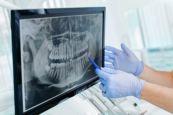
An Orthopantomogram (OPG) is a dental X-ray that captures a panoramic view of the entire oral and maxillofacial region. Unlike intraoral X-rays, which focus on specific areas, OPG provides a broader perspective, including:
- all teeth (upper and lower jaws);
- surrounding bone structures;
- temporomandibular joints (TMJ);
- sinuses.
This comprehensive image helps dentists assess the overall health of the oral cavity in a single scan, making it an efficient and effective diagnostic tool.
Why is OPG important?
OPG plays a critical role in detecting and diagnosing various dental and maxillofacial conditions. Here’s a closer look at some of the key uses:
1. Early detection of hidden issues
OPG allows for the identification of dental and jaw problems that might not be visible during a clinical examination. These include:
- Cyst, tumors, or bone abnormalities
Dental cysts, tumors, and other bone irregularities are often asymptomatic in their early stages. OPG helps detect these conditions early, allowing for timely intervention. Panoramic X-rays improve the early detection of bone pathologies that could otherwise go unnoticed.
- Dental impactions
Impacted teeth, especially wisdom teeth, can cause pain, infection, or misalignment. OPG imaging provides a clear view of their position, helping dentists determine whether extraction or monitoring is necessary. Panoramic imaging remains the gold standard for diagnosing and planning treatment for impacted third molars.
- Early signs of periodontal disease
Periodontal disease, commonly known as gum disease, is a progressive condition that affects the supporting structures of the teeth. Early detection is crucial to prevent irreversible damage. OPG plays a significant role in identifying early signs of periodontal disease by providing a panoramic view of the jawbone.
2. Comprehensive treatment planning
By offering a complete view of the teeth, jaws, and surrounding structures, OPG helps dentists plan treatments with greater accuracy. This is particularly important for:
- Orthodontics
For patients considering braces or clear aligners, OPG combined with Cephalometric offers a clear overview of tooth alignment and jaw structure, ensuring precise treatment planning. Dentists can identify underlying issues like crowding, impacted teeth or over jets that need to be addressed before orthodontic procedures. - Implant placement
OPG usage in implant planning, noting its effectiveness in assessing bone quality and anatomical considerations essential for implant success
- Wisdom tooth extraction
OPG enables clinicians to visualize the angulation, impaction status, and relationship of wisdom teeth to adjacent teeth and the inferior alveolar nerve.
This comprehensive view assists in determining the complexity of extraction and in formulating a surgical approach that minimizes risks such as nerve injury or postoperative complications.
3. Improved patient understanding
The panoramic nature of OPG image makes it easier for patients to visualize their dental conditions. Dentists can use the images to explain diagnoses and proposed treatments, fostering trust and understanding between patients and providers.
How does OPG work?
The OPG procedure is quick, painless, and efficient. Here’s what to expect during the process:
- Preparation: You will be asked to remove any jewelry, glasses, or metal objects near your face to prevent artefacts from the radiographed site. You will be asked to wear a lead apron during exposure.
- Positioning: You will stand while placing your chin on a support and biting on a sterile mouthpiece to keep your head stable.
- Imaging: The machine’s arm rotates around your head, capturing a detailed panoramic image. The process typically takes less than two minutes.
Common patient questions about OPG
1. Is OPG safe?
Yes, OPG is safe for most patients. The radiation dose is minimal, and protective measures are taken to ensure safety. Pregnant patients should inform their dentist to discuss alternative diagnostic options.
2. Do I need an OPG?
Your dentist may recommend an OPG if you are experiencing:
- planning orthodontic treatment;
- suspected wisdom tooth impaction;
- considering dental implants.
3. How often should I have an OPG?
OPG frequency depends on your oral health needs. For example, patients undergoing orthodontics or implant planning may need periodic scans, while others might only require it during comprehensive check-ups.
Conclusion
Dental Panoramic X-rays (OPG) are a cornerstone of modern dental diagnostics, offering unparalleled insight into oral and maxillofacial health. From detecting impacted teeth to planning complex treatments like implants, OPG ensures precision and safety at every step.
At GWS Medika Blok M Dental Clinic, our commitment to leveraging advanced technology like OPG ensures that your oral health is in expert hands.
Ready to take the next step in your dental care journey? Contact us today to schedule your OPG consultation!


